


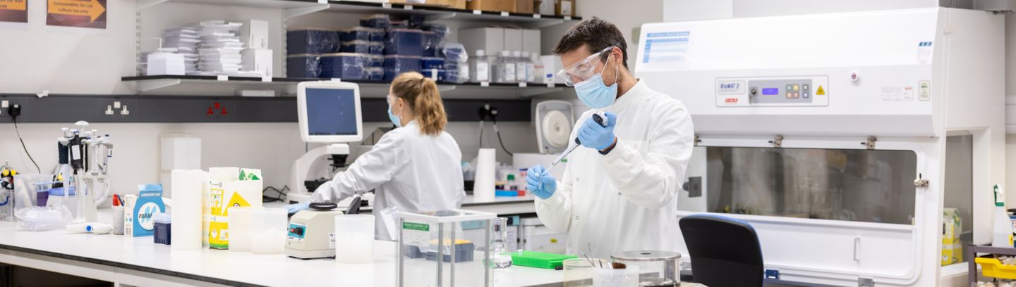
Postgraduate programmes for research careers
Top links
NHLI news
More news

NHLI Events
More events
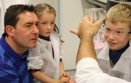
Engaging with the public and fellow researchers
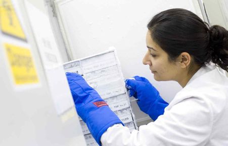
Improving cardiac and respiratory health through research and education
--tojpeg_1456231391004_x4.jpg)